上海大学学报(自然科学版) ›› 2022, Vol. 28 ›› Issue (5): 831-840.doi: 10.12066/j.issn.1007-2861.2442
窦涵钰, 崔白苹, 丁小雷( )
)
收稿日期:2022-07-27
出版日期:2022-10-30
发布日期:2022-11-12
通讯作者:
丁小雷
E-mail:xlding@shu.edu.cn
作者简介:丁小雷(1977—), 男, 教授, 研究方向为皮肤损伤修复及衰老分子机理、毛发再生、皮肤功能改善新药及新材料. E-mail: xlding@shu.edu.cn
DOU Hanyu, CUI Baiping, DING Xiaolei()
Received:2022-07-27
Online:2022-10-30
Published:2022-11-12
Contact:
DING Xiaolei
E-mail:xlding@shu.edu.cn
摘要:
皮肤是保护人体免受侵害的第一道防线, 常因接触物理、化学因子及微生物与病原体等造成各种创面. 创面的修复需要多种细胞、细胞因子和细胞外基质相互协同来完成. 由于过度炎症反应以及细胞外基质表达沉积与交联异常, 创面再生修复通常不能实现, 而是伴随着不同程度的瘢痕形成. 瘢痕的形成影响皮肤功能, 给患者带来心理障碍、身体痛苦, 严重影响生活质量, 但目前对瘢痕治疗尚缺乏特异且有效的药物. 近几年, 人们利用动物模型、单细胞分析、示踪成像等技术对于创面瘢痕形成的具体分子生物学机制进行了深入广泛研究. 讨论创面瘢痕形成机制的最新研究进展, 深入阐释瘢痕形成机制, 为未来研究抗疤痕药物和组织再生提供新思路.
中图分类号:
窦涵钰, 崔白苹, 丁小雷. 创面瘢痕形成机制研究进展[J]. 上海大学学报(自然科学版), 2022, 28(5): 831-840.
DOU Hanyu, CUI Baiping, DING Xiaolei. Advances in understanding the mechanism of wound scar formation[J]. Journal of Shanghai University(Natural Science Edition), 2022, 28(5): 831-840.
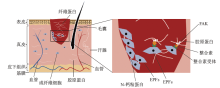
图1
成纤维细胞介导瘢痕形成过程"

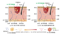
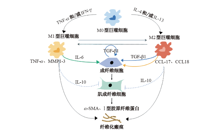
图2
巨噬细胞介导瘢痕形成过程"
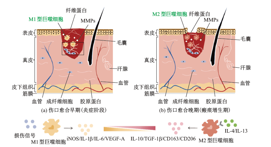
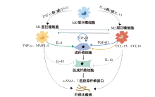
图3
瘢痕形成过程中成纤维细胞与巨噬细胞的相互作用"
| [1] |
Proksch E, Brandner J M, Jensen J M. The skin: an indispensable barrier[J]. Experimental dermatology, 2008, 17: 1063-1072.
doi: 10.1111/j.1600-0625.2008.00786.x pmid: 19043850 |
| [2] |
Rodrigues M, Kosaric N, Bonham C A, et al. Wound healing: a cellular perspective[J]. Physiological Reviews, 2019, 99: 665-706.
doi: 10.1152/physrev.00067.2017 pmid: 30475656 |
| [3] |
Sen C K, Gordillo G M, Roy S, et al. Human skin wounds: a major and snowballing threat to public health and the economy[J]. Wound Repair and Regeneration. 2009, 17: 763-771.
doi: 10.1111/j.1524-475X.2009.00543.x pmid: 19903300 |
| [4] |
Chesko D M, Wilgus T A. Immune cells in cutaneous wound healing: a review of functional data from animal models[J]. International Journal of Molecular Sciences, 2022, 23(5): 2044.
doi: 10.3390/ijms23042044 |
| [5] |
Feng Y, Sun Z L, Liu S Y, et al. Direct and indirect roles of macrophages in hypertrophic scar formation[J]. Frontiers in Physiology, 2019, 10: 1101.
doi: 10.3389/fphys.2019.01101 pmid: 31555142 |
| [6] |
Buechler M B, Fu W, Turley S J. Fibroblast-macrophage reciprocal interactions in health, fibrosis, and cancer[J]. Immunity, 2021, 54: 903-915.
doi: 10.1016/j.immuni.2021.04.021 pmid: 33979587 |
| [7] |
Longaker M T, Whitby D J, Adzick N S, et al. Studies in fetal wound healing, Ⅵ. Second and early third trimester fetal wounds demonstrate rapid collagen deposition without scar formation[J]. Journal of Pediatric Surgery, 1990, 25: 63-68.
pmid: 2299547 |
| [8] |
Marshall C D, Hu M S, Leavitt T, et al. Cutaneous scarring: basic science, current treatments, and future directions[J]. Advances in Wound Care, 2018, 7: 29-45.
doi: 10.1089/wound.2016.0696 pmid: 29392092 |
| [9] |
Rinkevich Y, Walmsley G G, Hu M S, et al. Skin fibrosis. Identification and isolation of a dermal lineage with intrinsic fibrogenic potential[J]. Science, 2015, 348: aaa2151.
doi: 10.1126/science.aaa2151 pmid: 25883361 |
| [10] |
Horsley V. Cut out that YAPping: mechanisms to reduce scar formation[J]. Cell Stem Cell, 2022, 29: 179-181.
doi: 10.1016/j.stem.2022.01.005 pmid: 35120615 |
| [11] |
Mascharak S, Des Jardins-Park H E, Davitt M F, et al. Preventing Engrailed-1 activation in fibroblasts yields wound regeneration without scarring[J]. Science, 2021, 372(6540): eaba2374.
doi: 10.1126/science.aba2374 |
| [12] |
Mascharak S, Talbott H E, Januszyk M, et al. Multi-omic analysis reveals divergent molecular events in scarring and regenerative wound healing[J]. Cell Stem Cell, 2022, 29: 315-327.
doi: 10.1016/j.stem.2021.12.011 pmid: 35077667 |
| [13] |
Philippeos C, Telerman S B, Oulès B, et al. Spatial and single-cell transcriptional profiling identifies functionally distinct human dermal fibroblast subpopulations[J]. The Journal of Investigative Dermatology, 2018, 138: 811-825.
doi: 10.1016/j.jid.2018.01.016 |
| [14] |
He H, Suryawanshi H, Morozov P, et al. Single-cell transcriptome analysis of human skin identifies novel fibroblast subpopulation and enrichment of immune subsets in atopic dermatitis[J]. The Journal of Allergy and Clinical Immunology, 2020, 145: 1615-1628.
doi: S0091-6749(20)30182-2 pmid: 32035984 |
| [15] |
Harper R A, Grove G. Human skin fibroblasts derived from papillary and reticular dermis: differences in growth potential in vitro[J]. Science, 1979, 204: 526-527.
pmid: 432659 |
| [16] |
Janson D G, Saintigny G, Van Adrichem A, et al. Different gene expression patterns in human papillary and reticular fibroblasts[J]. The Journal of Investigative Dermatology, 2012, 132: 2565-2572.
doi: 10.1038/jid.2012.192 |
| [17] |
Driskell R R, Lichtenberger B M, Hoste E, et al. Distinct fibroblast lineages determine dermal architecture in skin development and repair[J]. Nature, 2013, 504: 277-281.
doi: 10.1038/nature12783 |
| [18] |
Gauglitz G G, Korting H C, Pavicic T, et al. Hypertrophic scarring and keloids: pathomechanisms and current and emerging treatment strategies[J]. Molecular Medicine, 2011, 17: 113-125.
doi: 10.2119/molmed.2009.00153 pmid: 20927486 |
| [19] |
Gabbiani G, Ryan G B, Majne G. Presence of modified fibroblasts in granulation tissue and their possible role in wound contraction[J]. Experientia, 1971, 27: 549-550.
doi: 10.1007/BF02147594 pmid: 5132594 |
| [20] |
Hinz B. The role of myofibroblasts in wound healing[J]. Current Research in Translational Medicine, 2016, 64: 171-177.
doi: S2452-3186(16)30039-3 pmid: 27939455 |
| [21] |
Hinz B, Gabbiani G. Mechanisms of force generation and transmission by myofibroblasts[J]. Current Opinion in Biotechnology, 2003, 14: 538-546.
doi: 10.1016/j.copbio.2003.08.006 pmid: 14580586 |
| [22] |
Padmanabhan J, Maan Z N, Kwon S H, et al. In vivo models for the study of fibrosis[J]. Advances in Wound Care, 2019, 8: 645-654.
doi: 10.1089/wound.2018.0909 pmid: 31827979 |
| [23] |
Sakar M S, Eyckmans J, Pieters R, et al. Cellular forces and matrix assembly coordinate fibrous tissue repair[J]. Nature Communications, 2016, 7: 11036.
doi: 10.1038/ncomms11036 pmid: 26980715 |
| [24] |
Zhao X K, Cheng Y, Liang C M, et al. Focal adhesion kinase regulates fibroblast migration via integrin beta-1 and plays a central role in fibrosis[J]. Scientific Reports, 2016, 6: 19276.
doi: 10.1038/srep19276 |
| [25] |
Jiang D, Christ S, Correa-Gallegos D, et al. Injury triggers fascia fibroblast collective cell migration to drive scar formation through N-cadherin[J]. Nature Communications, 2020, 11: 5653.
doi: 10.1038/s41467-020-19425-1 pmid: 33159076 |
| [26] |
Takeichi M. Cadherins: a molecular family important in selective cell-cell adhesion[J]. Annual Review of Biochemistry, 1990, 59: 237-252.
pmid: 2197976 |
| [27] |
Wan L, Jiang D, Correa-Gallegos D, et al. Connexin 43 gap junction drives fascia mobilization and repair of deep skin wounds[J]. Matrix Biology : Journal of the International Society for Matrix Biology, 2021, 97: 58-71.
doi: 10.1016/j.matbio.2021.01.005 |
| [28] |
Kotini M, Barriga E H, Leslie J, et al. Gap junction protein Connexin-43 is a direct transcriptional regulator of N-cadherin in vivo[J]. Nature Communications, 2018, 9: 3846.
doi: 10.1038/s41467-018-06368-x pmid: 30242148 |
| [29] |
Li X, Guo L, Yang X, et al. TGF-$\beta $1-induced connexin43 promotes scar formation via the Erk/MMP-1/collagen Ⅲ pathway[J]. Journal of Oral Rehabilitation, 2020, 47: 99-106.
doi: 10.1111/joor.12829 |
| [30] |
Mosser D M, Edwards J P. Exploring the full spectrum of macrophage activation[J]. Nature Reviews Immunology, 2008, 8: 958-969.
doi: 10.1038/nri2448 pmid: 19029990 |
| [31] |
Wei S, Chow L T, Shum I O, et al. Left and right ventricular collagen type Ⅰ/Ⅲ ratios and remodeling post-myocardial infarction[J]. Journal of Cardiac Failure, 1999, 5: 117-126.
pmid: 10404351 |
| [32] |
Davies L C, Taylor P R. Tissue-resident macrophages: then and now[J]. Immunology, 2015, 144: 541-548.
doi: 10.1111/imm.12451 pmid: 25684236 |
| [33] |
Iwasaki A, Medzhitov R. Control of adaptive immunity by the innate immune system[J]. Nature Immunology, 2015, 16: 343-353.
doi: 10.1038/ni.3123 pmid: 25789684 |
| [34] |
Willenborg S, Lucas T, Van Loo G, et al. CCR2 recruits an inflammatory macrophage subpopulation critical for angiogenesis in tissue repair[J]. Blood, 2012, 120: 613-625.
doi: 10.1182/blood-2012-01-403386 pmid: 22577176 |
| [35] |
Hesketh M, Sahin K B, West Z E, et al. Macrophage phenotypes regulate scar formation and chronic wound healing[J]. International Journal of Molecular Sciences, 2017, 18(7): 1545.
doi: 10.3390/ijms18071545 |
| [36] |
Knipper J A, Willenborg S, Brinckmann J, et al. Interleukin-4 receptor $\alpha $ signaling in myeloid cells controls collagen fibril assembly in skin repair[J]. Immunity, 2015, 43: 803-816.
doi: 10.1016/j.immuni.2015.09.005 |
| [37] |
Sindrilaru A, Peters T, Wieschalka S, et al. An unrestrained proinflammatory M1 macrophage population induced by iron impairs wound healing in humans and mice[J]. The Journal of Clinical Investigation, 2011, 121: 985-997.
doi: 10.1172/JCI44490 |
| [38] |
Herold S, Mayer K, Lohmeyer J. Acute lung injury: how macrophages orchestrate resolution of inflammation and tissue repair[J]. Frontiers in Immunology, 2011, 2: 65.
doi: 10.3389/fimmu.2011.00065 pmid: 22566854 |
| [39] |
Khallou-Laschet J, Varthaman A, Fornasa G, et al. Macrophage plasticity in experimental atherosclerosis[J]. PLoS One, 2010, 5: e8852.
doi: 10.1371/journal.pone.0008852 |
| [40] |
Anderson-Baucum E, Piñeros A R, Kulkarni A, et al. Deoxyhypusine synthase promotes a pro-inflammatory macrophage phenotype[J]. Cell Metabolism, 2021, 33: 1883-1893.
doi: 10.1016/j.cmet.2021.08.003 pmid: 34496231 |
| [41] |
Kraakman M J, Murphy A J, Jandeleit-Dahm K, et al. Macrophage polarization in obesity and type 2 diabetes: weighing down our understanding of macrophage function?[J]. Frontiers in Immunology, 2014, 5: 470.
doi: 10.3389/fimmu.2014.00470 pmid: 25309549 |
| [42] |
Vannella K M, Wynn T A. Mechanisms of organ injury and repair by macrophages[J]. Annual Review of Physiology, 2017, 79: 593-617.
doi: 10.1146/annurev-physiol-022516-034356 pmid: 27959618 |
| [43] |
Das A, Sinha M, Datta S, et al. Monocyte and macrophage plasticity in tissue repair and regeneration[J]. The American Journal of Pathology, 2015, 185: 2596-2606.
doi: 10.1016/j.ajpath.2015.06.001 |
| [44] |
Zhu Y, Li X, Chen J, et al. The pentacyclic triterpene Lupeol switches M1 macrophages to M2 and ameliorates experimental inflammatory bowel disease[J]. International Immunopharmacology, 2016, 30: 74-84.
doi: S1567-5769(15)30199-5 pmid: 26655877 |
| [45] |
Chen L, Li Z, Zheng Y, et al. 3D-printed dermis-specific extracellular matrix mitigates scar contraction via inducing early angiogenesis and macrophage M2 polarization[J]. Bioactive Materials, 2022, 10: 236-246.
doi: 10.1016/j.bioactmat.2021.09.008 pmid: 34901542 |
| [46] |
Kurose H, Mangmool S. Myofibroblasts and inflammatory cells as players of cardiac fibrosis[J]. Archives of Pharmacal Research, 2016, 39: 1100-1113.
doi: 10.1007/s12272-016-0809-6 pmid: 27515051 |
| [47] |
Zhu Z, Ding J, Ma Z, et al. Systemic depletion of macrophages in the subacute phase of wound healing reduces hypertrophic scar formation[J]. Wound Repair and Regeneration, 2016, 24: 644-656.
doi: 10.1111/wrr.12442 pmid: 27169512 |
| [48] |
Funes S C, Rios M, Escobar-Vera J, et al. Implications of macrophage polarization in autoimmunity[J]. Immunology, 2018, 154: 186-195.
doi: 10.1111/imm.12910 pmid: 29455468 |
| [49] |
Goren I, Allmann N, Yogev N, et al. A transgenic mouse model of inducible macrophage depletion: effects of diphtheria toxin-driven lysozyme M-specific cell lineage ablation on wound inflammatory, angiogenic, and contractive processes[J]. The American Journal of Pathology, 2009, 175: 132-147.
doi: 10.2353/ajpath.2009.081002 |
| [50] |
Franz S, Ertel A, Engel K M, et al. Overexpression of S100A9 in obesity impairs macrophage differentiation via TLR4-NF$\kappa$B-signaling worsening inflammation and wound healing[J]. Theranostics, 2022, 12: 1659-1682.
doi: 10.7150/thno.67174 |
| [1] | 刘仲尼, 杨力明. 半乳糖凝集素-3 与巨噬细胞炎性反应的关系[J]. 上海大学学报(自然科学版), 2017, 23(3): 395-401. |
| [2] | 肖珍, 朱杰宁, 唐春梅, 林秋雄, 胡志琴, 张灼, 符永恒, 张梦珍, 单志新. 巨噬细胞移动抑制因子缺失加重苯肾上腺素诱导的小鼠心肌肥厚[J]. 上海大学学报(自然科学版), 2016, 22(3): 336-343. |
| 阅读次数 | ||||||
|
全文 |
|
|||||
|
摘要 |
|
|||||